The rate of attic cholesteatomas occurring behind a tiny retraction of the pars flaccida was 7 7 11 of 142 patients with attic cholesteatoma.
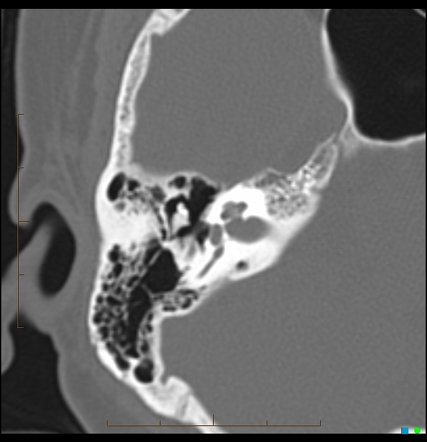
Attic retraction ct.
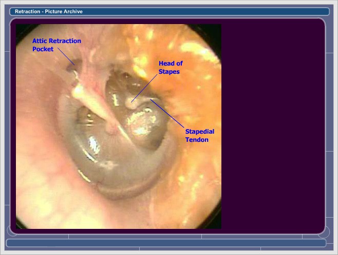
As the tympanic membrane is pulled inwards medially it can become draped over the ossicles resulting in a variety of symptoms.
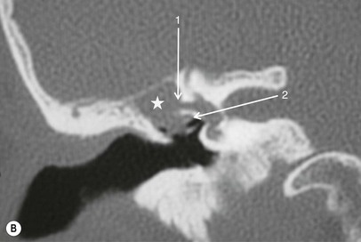
Cholesteatomas appear as regions of soft tissue attenuation exerting mass effect and resulting in bony erosion.
Tympanic membrane retraction usually occurs when a portion of the tympanic membrane becomes weakened and is pulled inwards by the negative pressure within the middle ear.
1 3 this entity results from a deep retraction pocket or a cholesteatoma that has eroded the bone and then spontaneously drained into the external auditory canal.
Ct is the modality of choice for diagnostic assessment of cholesteatomas due to its ability to demonstrate the bony anatomy of the temporal bone in exquisite detail.
An auto atticotomy also called nature s atticotomy refers to an enlarged lateral attic with absence of the scutum and lower lateral wall of the attic in a patient without a history of surgery.
Invagination of the tympanic membrane of the attic to form retraction pockets to be filled with desquamated epithelium and keratin to form cholesteatoma.
The pertinent anatomy is described and the role of the tympanic diaphragm and isthmus in determining the degree to which middle ear disease may progress is stressed.
Revision surgery is performed in cases of functional failure residual cholesteatoma suspected on follow up ct scan or mri and recurrent cholesteatoma.
Eustachian tube theory.
To examine this theory computerized tomographic ct findings of these conditions were.
Attic and the adjacent middle ear spaces and that subsequent building up of the negative pressure in these spaces results in retraction of the pars flaccida leading to formation of attic retraction pockets and cholesteatomas.
Management of controlled posterior or posterior attic retraction pockets.
Download citation a tiny retraction of the pars flaccida may conceal an attic cholesteatoma objective the purpose of this study was to evaluate the possibility of attic cholesteatomas.
This is the most common and widely considered as the main reason for cholesteatoma.
The retraction can be subdivided based on severity 1.
For further follow up an mri exam is performed once a year for 5 years especially in children.